A brain on a chip towards a cure for Parkinson’s disease
Experts
Coordinator
Patrik Verstreken (VIB-KU Leuven)
Partners
Dries Braeken (imec)
Birgitt Schuele (Stanford University School of Medicine)
Wim Vandenberghe (UZ Leuven)
Adrian Ranga (KU Leuven)
Magali Haas (Cohen Veterans Bioscience)
Why
The living human brain is difficult to study. Most detailed analyses can only be done post-mortem, thereby providing just a snapshot, and often in the end stages when we can no longer observe the processes that have triggered and driven disease. Moreover, current experimental models are useful but do not reliably reproduce all aspects of the human condition. We also lack an early diagnostic tool that would allow us to identify people who are developing brain disease before major damage has occurred.
How
Close collaboration between engineers, biomedical researchers and clinicians, supported by substantial investment from governments, funding agencies and philanthropists, can help us decode and ultimately cure Parkinson’s disease.
Our aim is for Parkinson’s to be objectively defined by solid molecular criteria, rather than by the typical movement symptoms that are currently used to diagnose the disease. This will mean scientists can develop targeted medical interventions that effectively slow down or even stop the disease based on molecular and genetic pathways.
We will develop new technology to create 2D and 3D “brain-on-a-chip” models. These will use patient skin cells to re-construct the neuronal networks of a living brain. The new tools will allow the stratification of patients and the testing of new, personalized therapeutic approaches. We can also apply them to other neurodegenerative diseases, including Amyotrophic Lateral Sclerosis (ALS) and Alzheimer’s.
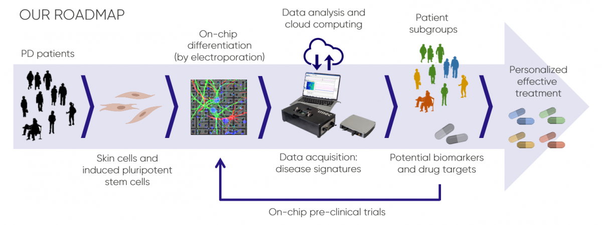
Imec’s advanced chip technologies will allow high-throughput interrogation of engineered three-dimensional neuronal circuits on a single-cell level. Bringing our platform technologies together with human stem cell lines from patients will aid in the development of better models for neurodegenerative diseases.
Partners
Publications
-
Brochure on the Brain on a Chip Project
Check out the flipbook of a new brochure that illustrates the challenges and goals of the team developing Brain on a Chip.
-
Duckert et al. 2022 (J Control Release) High-definition electroporation: Precise and efficient transfection on a microelectrode array.
This publication describes the experimental method developed in the lab of Dr. Dries Braeken to deliver small molecules into cells via on-chip electroporation. This technique can be used for various biomedical applications, including mRNA transfection and gene editing.
-
Mastrangeli et al. 2019 (ALTEX) Building blocks for a European Organ-on-Chip roadmap
In January 2019, Dries Braeken attended the Organ-on-Chip In Development (ORCHID) Strategy workshop which intended to establish a European Organ-on-Chip roadmap. This resulted in six building blocks, which are detailed in this report.
- Miccoli et al. 2019 (Front Neurosci) High-Density Electrical Recording and Impedance Imaging With a Multi-Modal CMOS Multi-Electrode Array Chip.
-
Miccoli et al. 2018 (Curr Pharm Des) Brain-on-a-chip Devices for Drug Screening and Disease Modeling Applications
This review discusses the current strategies to make brain-on-chip devices, their role in the study of the healthy and diseased brain, and their limitations and future perspectives.
-
Mora Lopez et al. 2018 (IEEE International Solid-State Circuits Conference on Feb 14, 2018) A 16384-Electrode 1024-Channel Multimodal CMOS MEA for High-Throughput Intracellular Action Potential Measurements and Impedance Spectroscopy in Drug-Screening Applications.
Researchers at imec have designed and fabricated a 16,384-electrode, 1,024-channel micro-electrode array (MEA) for high-throughput multi-modal cell interfacing. The chip offers intracellular and extracellular recording, voltage- and current-controlled stimulation, impedance monitoring and spectroscopy functionalities thereby packing the most cell-interfacing modalities on a single chip, and being the only one to enable multi-well assays. With this new chip, imec has created a platform that enables high quality data acquisition at increased throughput in cell-based cell studies. (Read more and watch the video)
-
Ju et al. 2017 (Nature) Fully integrated silicon probes for high-density recording of neural activity.
Engineers and scientists at imec, KU Leuven and VIB collaborated with researchers at HHMI’s Janelia Research Campus, the Allen Institute, and University College London (with grant funding from Gatsby and Wellcome) to build and test powerful new devices for detecting neural activity within the brains of living animals. The result is a silicon probe called Neuropixels, which can simultaneously record the activity of more than 200 individual neurons.
-
July 2017 | Chip technology for brain probes
Imec does not only develop multi-electrode probes to probe brain activity, but also probes that can stimulate the activity of brain cells by shining light on them. Such probes can be used for ‘optogenetics’, a technique combining genetics and optics.